
Human brain organoids have fundamentally shifted the paradigm of neurological research. By transforming human pluripotent stem cells (hPSCs) into self-organizing, three-dimensional tissues, these models provide an unprecedented window into the distinct spatial architecture and cellular diversity of the developing human brain. Unlike traditional two-dimensional cultures or animal models, which frequently fail to replicate human-specific evolutionary traits and disease phenotypes, 3D organoids allow scientists to observe complex neurodevelopmental processes directly in a human genetic background.
However, as the field transitions from initial discovery to translational application, a critical bottleneck has emerged: physiological relevance. Standard unguided organoids often suffer from significant batch-to-batch variation, a lack of structured non-ectodermal cell types, and a necrotic core caused by the absence of a functional vascular system. To unlock the full potential of these models for high-throughput drug screening and accurate disease modeling, researchers are increasingly turning to advanced bioengineering tools and region-specific differentiation protocols.
The Challenge of Reproducibility and Region-Specific Fidelity
One of the primary hurdles in organoid research is balancing self-organization with structural predictability. Early protocols relied heavily on untargeted intrinsic differentiation, which yielded heterogeneous tissues containing unpredictable mixtures of brain regions. For comparative disease modeling, this variance introduces confounding factors that can mask subtle pathological phenotypes.
To solve this, specialized protocols utilize precise chronological applications of small molecules and growth factors to steer cells toward specific lineages. This targeted approach has enabled the generation of discrete region-specific models, such as cortical, hippocampal, and midbrain organoids. For instance, normal midbrain organoid models can now be reliably differentiated to express a high density of functional dopaminergic neurons, providing a crucial baseline for studying neurodegenerative disorders like Parkinson’s disease.
Despite these advances, microenvironmental constraints still limit tissue longevity. In vivo, the brain relies on a dense capillary network to deliver oxygen and nutrients while clearing metabolic waste. In vitro, organoids depend entirely on passive diffusion. Once an organoid grows beyond a critical radius (typically 200 to 400 micrometers), its core suffers from hypoxia and subsequent cell death. This necrotic center disrupts physiological signaling and limits the organoid’s maturation, preventing it from progressing past embryonic developmental stages.
Advanced Bioengineering Solutions: Vascularization and Microfluidics
To overcome the diffusion limit and enhance structural fidelity, structural biologists and bioengineers are integrating advanced technological platforms into tissue culture workflows:
- Microfluidic Organ-on-a-Chip Systems: By housing organoids within specialized microfluidic chambers, researchers can introduce continuous, laminar fluid flow. This dynamic perfusion mimics the mechanical shear stress experienced by blood vessels in vivo, promoting enhanced cellular differentiation, faster functional maturation, and a significant reduction in core necrosis.
- Co-Culture and De Novo Vascularization: Another cutting-edge approach involves co-culturing hPSCs with endothelial cells or pericytes to induce the formation of primitive blood-vessel-like networks within the organoid. These primitive vessels can later be integrated with microfluidic channels to establish a fully pump-perfused vascular system.
- Bioprinting and Scaffolds: Utilizing biocompatible hydrogels and precise 3D bioprinting allows scientists to control the initial spatial distribution of cells, guiding the organoid into a more physiologically accurate anatomical shape from day one.
Beyond structural scaffolding, achieving true physiological relevance requires incorporating non-neuronal cell types that originate outside the neuroectoderm. For example, microglia—the resident immune cells of the central nervous system—arise from the embryonic yolk sac and play a vital role in synaptic pruning and neuroinflammation. Incorporating microglia into neural organoid models is essential for accurately mimicking neurodevelopmental and neuroinflammatory diseases, such as autism spectrum disorders and microcephaly.
Join the Discussion: Live Expert Insights
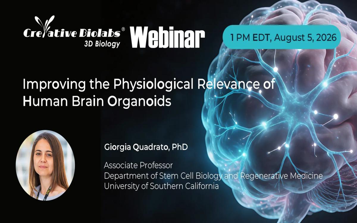
As these bioengineering methods mature, establishing standard operational procedures and scalable solutions remains crucial for industrial and academic research. To address these technical frontiers, Creative Biolabs is hosting a free, expert-led industry webinar titled “Improving the Physiological Relevance of Human Brain Organoids” on August 5, 2026, from 01:00 PM to 02:00 PM EDT, focusing on the latest methodologies driving organoid fidelity.
The session will feature Giorgia Quadrato, PhD, Associate Professor in the Department of Stem Cell Biology and Regenerative Medicine at the University of Southern California (USC) and Director of the USC CIRM ASCEND Shared Resource Laboratory. Dr. Quadrato’s pioneering research focus centers on human pluripotent stem cell differentiation, neurodevelopmental modeling, and the integration of novel bioengineering tools to study cell-type-specific mechanisms in developmental disease.
During the live presentation, Dr. Quadrato will break down strategic frameworks for generating highly reproducible, region-specific models, utilizing bioengineering interventions to sustain long-term organoid culture, and leveraging these systems to uncover novel disease mechanisms.
Translational Solutions for 3D Biology
Supporting these academic and clinical breakthroughs requires robust logistical infrastructure and validated biological systems. Specialized biotechnology providers, such as Creative Biolabs, have developed comprehensive platforms to streamline 3D tissue workflows. Through their dedicated 3D biology model gallery, the organization offers standardized biological tools designed to minimize experimental variance.
Their technical portfolio includes a fully customizable neural organoid model platform, which assists researchers in establishing stable, long-term cultures derived from validated human iPSC lines. For downstream applications, they supply specific targeted lineages, such as the normal midbrain organoid model, helping to eliminate the labor-intensive trial-and-error phase traditionally associated with home-brew differentiation protocols. By providing ready-to-use, quality-controlled 3D models alongside comprehensive contract research services, these platforms allow target validation and neurotoxicity screening teams to scale up their pipelines with higher confidence.
Ultimately, the fusion of developmental biology, microfluidic engineering, and standardized commercial solutions is accelerating the transition of brain organoids from structural novelties into rigorous, high-throughput discovery tools. Secure your complimentary registration for the upcoming webinar to stay at the absolute forefront of these next-generation neurological models.
Author Bio
Dr. Emily R. Coleman is a senior scientist at Creative Biolabs with a background in immunology, oncology research, and translational biotherapeutic development. Her work focuses on translating complex biological mechanisms into practical experimental strategies for next-generation therapeutic discovery, spanning both immune system biology and disease modeling platforms.
In addition to her core expertise in tumor immunology and antibody engineering, Dr. Coleman has contributed to cross-disciplinary research initiatives involving neurodegenerative disease modeling and human cell-based assay development. Her recent work includes supporting integrated preclinical research strategies that leverage advanced in vitro systems, including human-derived cellular platforms relevant to neurodegeneration research.
At Creative Biolabs, she provides scientific insight across multiple R&D domains, including antibody discovery and development, gene and cell therapy research, and translational assay design. She is particularly focused on improving the connection between mechanistic biology and predictive preclinical models to support more effective therapeutic development across complex disease areas, including neurodegenerative disorders and immune-related diseases.