New research suggests that inflammatory and microstructural changes in a key brain reward region may differ between chronic depression and acute depressive symptoms, offering fresh insight into the biological mechanisms underlying the condition.
In a large scale imaging study using data from the UK Biobank, researchers identified distinct magnetic resonance imaging markers in the ventral tegmental area, a dopamine rich brain region involved in motivation and reward processing, that were associated both with a history of major depressive disorder and with current symptom severity.
The findings, published in Biological Psychiatry: Cognitive Neuroscience and Neuroimaging, highlight the ventral tegmental area as a potential site where inflammatory processes and structural changes intersect in depression.
Investigating inflammation in the brain’s reward circuitry
Depression affects an estimated 300 million people worldwide and is increasingly understood as a multifactorial disorder in which inflammation plays an important role. Previous research has shown elevated inflammatory markers in blood and cerebrospinal fluid in people with depression, as well as altered dopamine signalling in reward related brain networks.
The ventral tegmental area sits at the centre of this network, regulating motivation, pleasure and mood through dopaminergic signalling. Despite its importance, the region has been difficult to study in detail because of its small size and complex anatomy.
In this study, researchers used advanced magnetic resonance imaging techniques to examine whether imaging derived markers sensitive to neuroinflammation and microstructure in the ventral tegmental area were associated with depression diagnosis and symptom severity.
Large scale analysis using UK Biobank data
The analysis included 32,495 participants from the UK Biobank who had diffusion weighted imaging, quantitative susceptibility mapping and structural MRI scans available. Of these, 3,807 participants had a history of major depressive disorder based on ICD 10 diagnosis codes. A matched group of healthy control participants was selected based on age, sex and body mass index.
To distinguish between long term depression history and current symptom burden, the researchers also used the Recent Depressive Symptoms questionnaire, which measures depressive symptoms experienced in the two weeks prior to scanning.
MRI metrics extracted from the ventral tegmental area included free water and isotropic volume fraction, which are sensitive to extracellular processes such as inflammation, as well as intracellular volume fraction and orientation dispersion index, which reflect microstructural properties. Magnetic susceptibility was also measured as an indirect marker of iron accumulation associated with chronic inflammation.
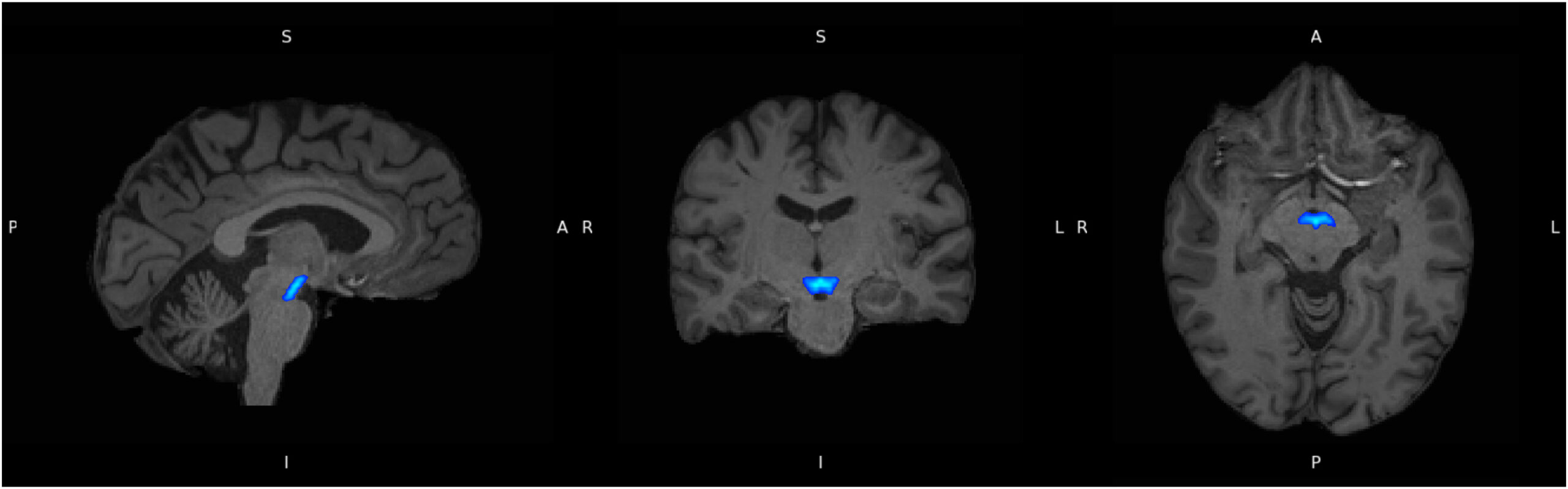
Ventral tegmental area (in blue), delineated using the Levinson-Bari Limbic Brainstem Atlas, displayed on coregistered participant T1 images. A, anterior; I, inferior; L, left; P, posterior; R, right; S, superior.
Distinct patterns for depression history and current symptoms
Participants with a history of major depressive disorder showed significantly higher free water and isotropic volume fraction in the ventral tegmental area compared with healthy controls. These findings indicate increased extracellular water content, consistent with inflammation related processes in this region.
In contrast, when the researchers examined predictors of current depressive symptom severity across the population, a different pattern emerged. Higher symptom severity was associated with increased intracellular volume fraction and orientation dispersion index, alongside lower isotropic volume fraction. These associations were independent of whether participants had a prior diagnosis of major depressive disorder.
Notably, magnetic susceptibility did not differ between groups and did not predict symptom severity, suggesting an absence of chronic inflammation related changes such as iron accumulation or cell damage in the ventral tegmental area.
The authors interpret these findings as evidence that acute depressive symptoms and long standing depression history may reflect distinct biological processes. Elevated extracellular water markers may indicate chronic inflammatory states, while changes in microstructural markers may reflect more dynamic or acute inflammatory responses.
Broader relevance across the brain
Exploratory analyses in other brain regions implicated in depression, including the hippocampus, amygdala, insula and orbitofrontal cortex, revealed similar increases in free water and isotropic volume fraction in individuals with a history of depression. This suggests that inflammation related changes are not confined to the ventral tegmental area but extend across reward and emotion related networks.
However, the ventral tegmental area appeared to show a unique pattern in relation to symptom severity, reinforcing its central role in the neurobiology of depression.
Limitations and future directions
The researchers note that the study is cross sectional and therefore cannot establish causal relationships between inflammation, brain structure and depressive symptoms. In addition, while MRI derived markers provide indirect evidence of inflammation, they do not directly measure immune activity. Longitudinal studies and integration with other biomarkers such as positron emission tomography or inflammatory cytokine measures will be needed to further clarify mechanisms.
Effect sizes were modest, reflecting the complex and multifactorial nature of depression, which is influenced by genetic, metabolic, lifestyle and psychosocial factors alongside neurobiological processes.
Implications for understanding depression
Despite these limitations, the study provides new evidence that inflammation related brain changes may differ between acute and chronic forms of depression. By distinguishing between markers associated with long term diagnosis and those linked to current symptom severity, the findings suggest that depression may not represent a single biological state but a dynamic process involving different inflammatory and structural patterns over time.
The authors conclude that these insights could inform more targeted therapeutic approaches that take into account whether depressive symptoms are acute or part of a longer term disorder, potentially improving treatment strategies in the future
Read the full paper
Khalife S, Bollmann S, Zalesky A, Oestreich LKL. Magnetic resonance imaging derived markers of acute and chronic inflammatory processes in the ventral tegmental area associated with depression. Biological Psychiatry: Cognitive Neuroscience and Neuroimaging. 2025. https://www.sciencedirect.com/science/article/pii/S2451902225002678?via%3Dihub
Feature Image – Cellular changes in response to varying degrees of inflammation. The figure illustrates the progression of neuroinflammation from a healthy state to acute and chronic inflammation