
Image duplication, one of the most common image integrity problems in published research, can result from an inadvertent mix-up during figure preparation, reuse that has not been clearly explained, or deliberate fabrication.
A 2025 analysis of the Retraction Watch database, covering more than 56,000 records, found around 8,000 retractions that cited images as a reason. Among the closely examined entries, the dominant pattern was duplication, most often the same image appearing in more than one paper [1]. Duplication is also one of the easiest mistakes to make, whether by an author, a team member or a collaborator.
That matters, because honest errors are also the easiest to prevent. An automated image integrity check before submission can catch them while they are still easy to fix. However, once a paper is published, the same error can lead to a correction or a retraction, with lasting consequences for the careers of everyone listed as an author on it. Not every result that fails to replicate comes from a flawed experiment. Sometimes the experiment was sound and the figure was presented incorrectly: the wrong panel chosen during assembly, a control image reused across two conditions, two overlapping fields recorded as independent replicates.
I spent years at the bench before working on this problem full time, including postdoctoral research in virology and immunology at Harvard Medical School and Beth Israel Deaconess Medical Center. What I noticed then, and have seen many times since, is how differently text and images are treated in review. Manuscript text is routinely checked through plagiarism screening, internal review and peer review. Images get far less scrutiny. And when someone does look at them, many problems simply cannot be seen with the naked eye.
How far one image travels
A micrograph captured to answer an internal question rarely stays there. It moves into a review deck, a preclinical data package, a partner data room, a manuscript, and in some cases a regulatory submission. Along the way it gets copied, cropped, relabeled and passed to new people.
Once published, it may be cited in a review, relied on in a funding application and built on in the next study. Eventually it can become part of the case for testing a new therapy in patients. Each person along that chain assumes someone earlier checked it.
Where the problems begin
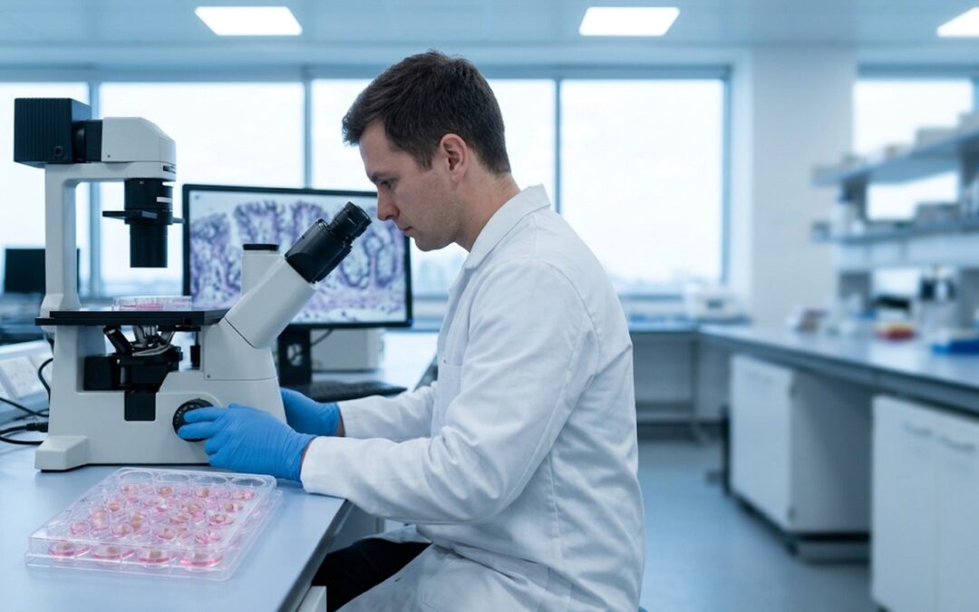
Take a typical microscopy figure. A student in the lab captures the images over several hours, moving the stage across the slide and changing magnification as they go. The instrument does not warn the user when fields overlap. Files are stored under names that made sense at the time. Eighteen months later that student has moved on, and a colleague assembles the panels from a shared drive, choosing representative images for research they did not personally run.
Preclinical and clinical work is frequently outsourced, split across contract research organizations and trial sites, then reassembled by people who were not present at capture. People leave, and work changes hands. Usually no one means any harm, but each of those steps is another chance for the wrong image to slip in.
How common are image integrity problems?
Image fabrication happens and it does real damage but it is not the only way a figure goes wrong. One image integrity analyst working across a set of journals reported that between 20 and 35 percent of manuscripts checked at acceptance were flagged for image-related problems [2]. Given how much imagery is produced on deadline by teams whose members keep changing, that statistic is not surprising.
Checking figures is not an accusation, especially given few manuscripts are the work of one person. Figures move between collaborators, students, contract laboratories and co-authors, and an error can enter at any hand-off. Yet every author named on a paper shares responsibility for the whole paper, however many hands the figures passed through. If a problem surfaces after publication, it is the authors, and above all the lead and corresponding authors, whose reputations are on the line. Verifying images before submission is part of that responsibility, and it is the most practical way for authors to protect their reputations.
What the eye cannot do
The human eye is simply not built for this kind of comparison. Nobody can keep hundreds of images in mind and check every one against all the others. Many scientific images offer the eye very little to focus on: one microscopy field or gel lane looks much like the next. That is just within one manuscript. Checking figures against the published literature, to confirm that no image has been reused from elsewhere, involves comparisons against millions of published images. A cross-sectional study published in 2025 tested medical students and researchers on spotting duplications in figures from published articles and found that they performed poorly [3]. The same consistency that makes an image good evidence makes its problems hard to spot by eye.
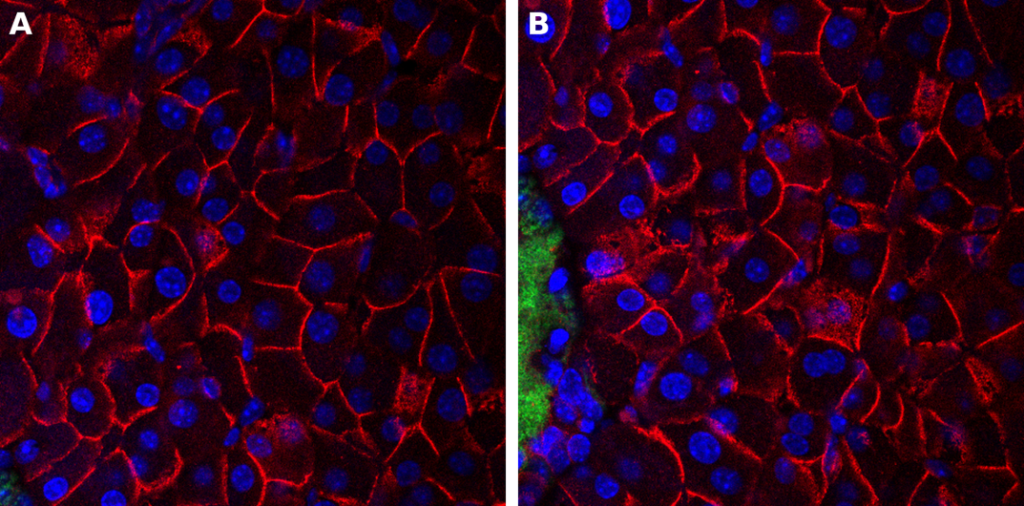
Can you spot the duplication? Two fluorescence micrographs of pancreatic tissue are shown as apparently different fields. A partially overlapping region is present in both panels, creating an inappropriate duplication that is difficult to detect by visual inspection alone. Image supplied by the author.
AI-generated images make this harder still. A duplicated image can be caught because a second copy exists somewhere. A synthetic western blot or micrograph has no second instance to compare against and no capture artifact to notice. A 2024 study published in Scientific Reports, with more than 800 participants, found that even those trained in histology could not reliably separate generated tissue samples from authentic ones [4]. A Nature news feature also highlighted this challenge [5].
The accountability gap, and how to close it
Figures are one of the few parts of a manuscript that often go to submission without being verified. The risk from that gap falls hardest on the authors who sign off.
Here are two practices that can help close that gap:
- Retain and organize originals, label files clearly, and record processing. From the outset, preserve unmodified original images with consistent filenames and clear links to the relevant experiments, samples, and conditions. Record any processing steps so that each figure panel can be traced back to its source. Uniform adjustments may be acceptable provided they do not obscure or distort information; selectively altering individual bands is not. The guidance established two decades ago remains relevant [6]. If questions arise years later, organized originals and a clear processing history help establish what happened and support a transparent, timely resolution.
- Verify with automated screening before submission. For image-heavy work, do not count on catching problems by eye. Build automated image verification into the workflow at each stage: when images are first collected from an experiment, before any analysis; before submission; and every time the manuscript is revised.
What automated screening can and cannot establish
A growing number of journals now build automated image screening into editorial review. The main reason is scale, not suspicion. A figure set of 100 sub-images generates several thousand unique pairs, each assessed across rotations, reflections and changes in magnification. No reviewer can work through that many comparisons reliably.
Automated image screening can flag suspicious similarities between image regions or signs that an image may have been generated rather than captured. It cannot establish intent. A flag is a reason to look closer, not a conclusion. Determining whether a flagged issue reflects an assembly error, legitimate reuse or deliberate manipulation requires clarification by the authors and assessment by the editors.
Catching problems earlier
A lot of effort now goes into finding image problems after publication. That work matters, but it is the most expensive time to intervene, and it often comes long after others have acted on the evidence. The correction itself is rarely the biggest cost. The bigger cost is everything that was built on the figure in the meantime.
The answer begins with earlier scrutiny, treated as routine quality control rather than an investigation, without prejudging the cause of a problem. Keeping original images clearly labeled, systematically organized and linked to their experiments from the outset, then combining automated screening with careful visual review before submission, are simple but crucial steps. Together, they help authors and editors resolve concerns before publication, protecting researchers, journals and institutions, as well as the future research that builds on their findings.
Author Bio