
A child’s vision is one of their most important developmental assets. Sight shapes how children learn, play, read, and connect with the world around them, and unlike many other aspects of health, vision loss caused by pediatric eye injuries can have consequences that follow a child for life.
Eye injuries are one of the leading causes of preventable vision loss in children [1]. In a U.S. emergency-department study of activity- and consumer-product-related pediatric eye injuries, children age 4 and younger accounted for 32% of cases and had the highest mean annual eye injury rate, at 11.31 per 10,000 children [8].
What makes this important is that children’s eyes are not fully developed. Early childhood is a critical window for visual development, and an injury during this period can disrupt the brain’s ability to build the pathways that support clear sight, sometimes leading to amblyopia, commonly known as lazy eye, even after the injury itself has healed [3].
The severity of an eye injury cannot always be judged by appearance alone. Some injuries appear alarming but turn out to be minor and heal fully with the right care. Others look deceptively mild: a child may seem comfortable and the eye may appear intact despite damage serious enough to threaten permanent vision loss. Knowing which is which, and when to refer, is what this article is about.

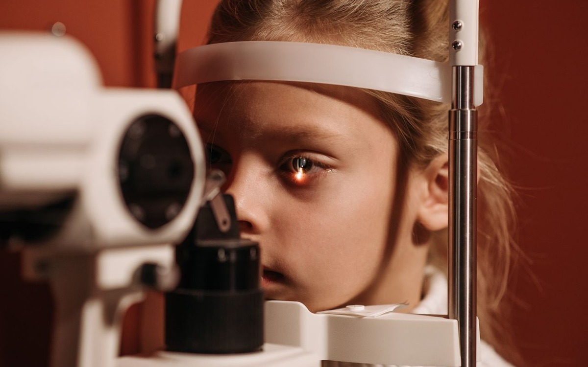
Image: Ivan S / Pexels
Eye Injury Types and Their Risk of Permanent Vision Loss
Not all eye injuries carry the same risk. The following table summarizes the major categories of pediatric eye injuries, their association with permanent vision loss, and the urgency of care required [3–7,11].
| Injury Type | Definition | Risk of Permanent Vision Loss | Requires Immediate Care? |
| Open globe / penetrating injury | A wound that breaks through the outer wall of the eye | High: corneal scarring, retinal damage, infection | Yes: emergency assessment |
| Chemical burn (alkali) | Damage from contact with basic substances like bleach or ammonia | Very high: rapid tissue destruction, limbal stem cell damage | Yes: immediate irrigation and specialist assessment |
| Retinal detachment (blunt trauma) | The retina pulls away from the back wall of the eye after a blow | High if untreated; lower with early surgery | Yes: emergency assessment for suspected detachment |
| Hyphema (blood in anterior chamber) | Bleeding into the front chamber of the eye between the cornea and iris | Moderate: risk of secondary glaucoma | Yes: same-day ophthalmology assessment |
| Traumatic cataract | The lens of the eye becomes clouded following injury | High risk of amblyopia in children if untreated | Prompt pediatric referral if the visual axis is affected |
| Traumatic optic neuropathy | Damage to the nerve that carries visual signals from the eye to the brain | High: nerve damage may be irreversible | Yes: urgent ophthalmology assessment |
| Corneal abrasion (minor) | A scratch on the clear front surface of the eye | Low, with proper treatment | Primary or urgent care assessment; refer for red flags |
| Orbital fracture | A break in the bones surrounding the eye socket | Depends on associated ocular injury; muscle entrapment needs urgent care | Urgent if diplopia, restricted motility, or vision change |
Clinical Features of High-Risk Pediatric Eye Injuries
Open Globe and Penetrating Injuries
An open globe injury means something has broken through the outer wall of the eye. This is one of the most serious eye injuries a child can experience.
Penetrating injuries can occur with everyday objects. Common examples include a stick or branch during outdoor play, a pencil or pen during a fall, a nail or screw in a home workshop, scissors during a craft activity, and broken glass or a sharp edge from a toy.
Once the eye wall is breached, the risk of infection, internal bleeding, and permanent damage to the retina or optic nerve increases significantly. How well the eye recovers depends on where the injury occurred, how deep it goes, and how quickly the child receives surgical care [3].
Immediate management: Do not apply pressure to the eye, perform tonometry, or attempt to remove an embedded object. Apply a rigid eye shield without pressure and arrange emergency ophthalmology consultation or transfer. A small or self-sealing wound may conceal an open globe injury; an irregular pupil, abnormal anterior chamber depth, or extensive subconjunctival hemorrhage should raise suspicion [3,11].
Chemical Burns
When a chemical splashes into a child’s eye, the damage begins within seconds, often before there is any visible sign of injury. Burns caused by alkali substances, such as bleach, drain cleaners, ammonia, and lime, are especially dangerous because they penetrate the eye rapidly and can continue to destroy tissue for hours after the initial contact. Acid burns, from substances like pool chemicals or battery acid, are also serious but tend to cause more contained damage.
In severe cases, chemical burns can cloud the cornea, cause chronic dry eye, and result in scarring that significantly reduces a child’s vision. Some children require a corneal transplant [4].
Immediate management: Begin irrigation immediately with saline or lactated Ringer solution if available, or clean water. Do not delay irrigation while obtaining a history or arranging referral. Continue until the ocular surface returns to near-physiologic pH, then recheck pH after stopping. Examine for retained particulate material and discuss the injury with ophthalmology [4,11].
Retinal Detachment After Blunt Trauma
The retina is the light-sensitive layer at the back of the eye that sends visual information to the brain. A forceful impact to the eye, such as from a ball, fist, or fall, can cause the retina to detach from the underlying tissue that supplies it with nutrients and support. Without surgery, permanent vision loss is likely. Even with prompt treatment, full recovery is not always possible, particularly if the central part of the retina is affected [5].
This type of injury is well documented in children who play contact sports or sports involving fast-moving projectiles, such as hockey, squash, and paintball.
Immediate management: New flashes, an increase in floaters, or a shadow or curtain across vision after trauma require urgent ophthalmic evaluation for a possible retinal tear or detachment. Suspected detachment is an emergency. Include these warning signs in discharge instructions because symptoms may develop after the first assessment [5,11].
Traumatic Cataract and Amblyopia
A hard blow or penetrating injury can cloud the lens of the eye, a condition called a traumatic cataract. In adults, this mainly causes blurred vision. In young children, the consequences can be more lasting. A clouded lens blocks the clear visual input the developing brain needs, and over time the brain may begin to ignore signals from that eye altogether, a condition called amblyopia, or lazy eye. This can persist even after the cataract is removed, particularly if treatment is delayed [3].
Immediate management: Significant visual-axis obstruction in a young child warrants prompt pediatric ophthalmology referral. Follow-up must address optical correction and amblyopia treatment as well as the cataract itself, because restoring the injured structure does not necessarily restore normal visual development [3].
Hyphema
Hyphema refers to bleeding within the eye, specifically into the space between the cornea (the clear, front surface) and the iris (the colored part of the eye). A blunt blow to the eye usually causes hyphema and a visible red layer at the bottom of the eye. Many hyphemas resolve with rest and medication, but two complications require careful monitoring: a second bleed and a dangerous rise in eye pressure that can damage the optic nerve over time. In a prospective series of 371 patients with traumatic hyphema, secondary hemorrhage occurred in 8% overall and in 5% of patients without identified risk factors. The cohort was not exclusively pediatric [6].
Immediate management: A child with a visible traumatic hyphema needs same-day ophthalmology assessment. Activity restrictions and follow-up should be directed by the treating ophthalmologist to monitor rebleeding and intraocular pressure [6,11].
Traumatic Optic Neuropathy
The optic nerve carries visual information from the eye to the brain. In severe trauma such as a serious car accident, a fall from height, or a significant sports injury, this nerve can be damaged directly or indirectly. Unlike most parts of the eye, the optic nerve has very limited ability to heal. The result can be permanent vision loss even when the eye itself looks completely normal from the outside [7].
Immediate management: Unexplained reduction in vision, impaired color vision, visual-field abnormalities, or a relative afferent pupillary defect after head or facial trauma warrants urgent ophthalmic assessment, even when the globe appears normal [7].
When Should a Child Be Referred After an Eye Injury?
One of the most clinically important points in pediatric eye injury management is this: clinicians should not determine severity from appearance alone. The following table provides guidance on referral and initial management [3–7,11]:
| Symptom or Sign | Initial Management or Referral |
| Sudden vision loss or significant blurring | Arrange emergency ophthalmic evaluation |
| Visible blood inside the eye (hyphema) | Arrange same-day ophthalmology assessment |
| Something stuck in or penetrating the eye | Do not remove; shield without pressure and arrange emergency consultation or transfer |
| Chemical splash or burn | Begin irrigation immediately; continue to near-physiologic pH and arrange urgent assessment |
| Eye appears misshapen, sunken, or has visible laceration | Suspect open globe injury; avoid pressure and arrange emergency consultation |
| Flashes of light or new floaters after injury | Urgent ophthalmic assessment for possible retinal tear or detachment |
| Pain that worsens after initial injury | Reassess and seek ophthalmology advice for deterioration |
| Persistent redness or light sensitivity 24 hours after injury | Reassess; refer if symptoms persist or the examination is abnormal |
| Minor redness or irritation, no vision change | Primary or urgent care assessment; follow up and refer for deterioration or delayed recovery |
An uncomplicated corneal abrasion can often be managed initially in primary or urgent care when visual acuity is preserved and penetrating injury, infection, and other red flags have been excluded. Persistent pain, worsening symptoms, or reduced vision requires reassessment and possible ophthalmology referral [11].
Prevention: Injury Evidence and Protective Eyewear Standards
Prevent Blindness estimates that appropriate safety eyewear can prevent 90% of eye accidents [2]. For sports specifically, the National Eye Institute states that appropriate protection can prevent nine out of ten sports-related eye injuries [9]. Prevention efforts should address the hazards identified in injury data and the performance of protective equipment.
1. Use Injury Data to Guide Prevention
A national study of activity- and consumer-product-related pediatric eye injuries treated in U.S. emergency departments from 1990–2009 found that 69% occurred at home. Sports and recreation accounted for 24%, and chemicals for 17% [8]. These findings support prevention beyond organized sports, including home and recreational exposures.
2. Match Protective Eyewear to the Hazard
The joint American Academy of Pediatrics and American Academy of Ophthalmology statement recommended polycarbonate sports eye protectors meeting applicable standards [10]. The AAP retired that policy in 2019. Current equipment selection should use the applicable sport-specific standard; ASTM F803-25 specifies performance requirements for eye protectors used in selected sports [12]. Ordinary eyeglasses do not provide equivalent protection [9].

Image: Tima Miroshnichenko / Pexels
3. Address Chemical Exposures
Chemical exposure accounted for 17% of injuries in the cited pediatric emergency-department study [8]. For clinical teams, prevention counseling should accompany clear instructions for immediate irrigation and access to urgent assessment after exposure [4,11].
4. Support Sports Safety Policies
Clinicians can use sports-safety counseling to explain the difference between ordinary eyewear and protective equipment designed for the activity. Schools, sports programs, and equipment manufacturers can use protective-eyewear standards when selecting and evaluating equipment [9,12].
5. Monitor Outcomes After Injury
Follow-up after pediatric trauma should assess both injury-related changes and visual development. Traumatic cataract, corneal scarring, and prolonged unequal visual input can require visual rehabilitation beyond anatomical repair [3].
Conclusion
Children’s eye injuries span a wide spectrum, from minor corneal abrasions that heal within days to open globe injuries and chemical burns that carry a substantial risk of permanent vision loss. What connects them is the critical importance of timely evaluation and treatment [3,11].
The younger the child, the higher the stakes. Because the visual system is still developing, even an injury that might be manageable in an adult can have lasting developmental consequences in a child. Early intervention for serious injuries and timely visual rehabilitation help preserve both vision and visual development [3,11].
For first-contact clinicians, the mechanism of injury, examination findings, visual function, and age of the child should guide referral urgency. An apparently intact eye does not exclude serious injury, and follow-up should account for delayed retinal symptoms and amblyopia risk [3,5,11].
Frequently Asked Questions
Which eye injuries in children are most likely to cause permanent vision loss?
Open globe and penetrating injuries, chemical burns (especially alkali), traumatic optic neuropathy, and untreated retinal detachments carry the highest risk of permanent vision loss. Traumatic cataracts in young children are also high-risk because of the associated risk of amblyopia during the critical developmental window [3–7].
Which findings require emergency assessment after an eye injury?
Arrange emergency assessment for sudden vision loss, an embedded object, a suspected open globe injury, or a visible wound or deformity of the eye. Chemical exposure requires immediate irrigation while urgent care is arranged. A visible traumatic hyphema requires a same-day ophthalmology assessment. Persistent redness, light sensitivity, or worsening pain should prompt reassessment [3–6,11].
Can a child’s eye injury cause amblyopia (lazy eye)?
Yes. If a traumatic cataract, corneal scar, or any other injury-related obstruction prevents clear images from reaching the retina during the developmental years, the brain may suppress input from that eye, leading to amblyopia. Prompt treatment and visual rehabilitation are especially important in young children [3].
What is the first step after chemical exposure to the eye?
Begin irrigation immediately with saline, lactated Ringer solution, or clean water. Continue until near-physiologic ocular surface pH is restored, recheck pH after stopping, and assess for retained material. Do not delay irrigation while arranging ophthalmology assessment [4,11].
Are all children’s eye injuries serious?
No. Many pediatric eye injuries, such as minor corneal abrasions from dust or debris, are superficial and heal fully with appropriate care. The challenge is distinguishing an uncomplicated abrasion from serious injury at assessment. Preserved vision and the absence of penetrating injury or other red flags support initial management in primary or urgent care; deterioration or delayed recovery warrants referral [11].
Author Bio